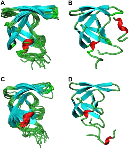
FIGURE 7.
The superposition of the 10 HIV-1 PR monomer conformations corresponding to the MD snapshots after each 1 ns during 10-ns simulation of the folding-binding mode (A) and dimerization-binding mode (C). The average structure of the peptide complex with the HIV-1 PR monomer obtained from 10-ns MD trajectory in the folding mode of inhibition (B) and dimerization mode of inhibition (D).

