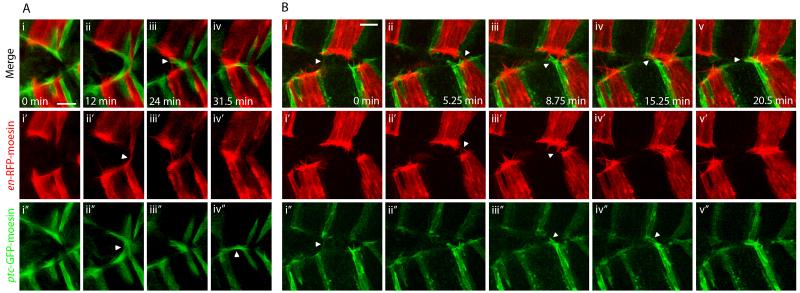
Figure 2. Cell matching and realignment during DC is mediated by filopodia.
Zippering in en-RFP-moesin (red), ptc-GFP-moesin (green) expressing embryos. A. Images from movie S2 showing filopodial matching. i. Red and green filopodia protrude from leading edge cells. ii. Contacts are made between red filopodia from opposing epithelia, while at the same time separate contacts are made between green filopodia. iii. Further contacts are made between red filopodia; however, green filopodia in close proximity to these red filopodial contacts do not interact. iii. Green filopodia transiently form contacts between ptc-GFP-moesin cells over the top of the fused red cells. B. Images from movie S3 showing realignment of misaligned epithelial sheets by filopodial searching and pulling. i. The two epithelial sheets are initially poorly aligned. A filopodial tether transiently exists between green cells but later breaks. ii. Contacts form between filopodia from en-RFP-moesin expressing cells. iii-iv. Green filopodia in the lower sheet do not interact with nearby red filopodia and ultimately form contacts with green cells some distance away in the opposing sheet. v. The tethers that result from the contacts made by red and green filopodia pull the sheets into alignment. Panels labelled ′ and ″ show en-RFP-moesin and ptc-GFP-moesin channels respectively in isolation. The described filopodial interactions are indicated by arrowheads. Scale bars 10 μm.

