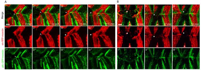
Figure 3. Matching in embryos with asymmetries between the opposing epithelial sheets.
DC zippering in en-RFP-moesin (red), ptc-GFP-moesin (green) embryos with asymmetries. A. Images from movie S4 showing zippering in an embryo with a spontaneous asymmetry. i. The upper epithelial sheet has two rather than one misplaced ptc cell in the en domain and one of these has associated with the A compartment of the lower sheet blocking matching of en cells. ii. As a result, the blocked en cells make filopodial contacts with en cells of the neighbouring segment. iii. These develop into permanent contacts. iv. Cells which are not associated with matching partners continue to produce filopodia. B. Zippering in an embryo with a laser-induced asymmetry. i. An en stripe has been removed from the leading edge of the lower epithelial sheet by laser ablation (indicated by asterisk) while the opposing en stripe (indicated by arrow) is normal. ii-iii. On contact with the opposing leading edge, the unpartnered en stripe constricts and then withdraws completely from the leading edge. Panels labelled ′ and ″ show en-RFP-moesin and ptc-GFP-moesin channels respectively in isolation. Scale bars 10 μm.

