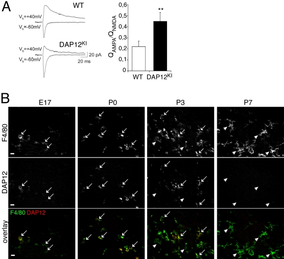
Figure 1. DAP12 mutation induces an increased ratio of AMPAR vs. NMDAR currents in adult hippocampal slices, whereas DAP12 is expressed only by microglia around birth.
(A) Ratio of AMPAR vs. NMDAR currents in hippocampal slices. Left, original traces from individual experiments. Right, averaged data from 9 and 10 recordings of WT and DAP12KI mice, respectively, Mean±SEM, **p<0.03, Mann-Whitney t test. (B) Double detection of DAP12 (upper line), of the microglial marker F4-80 (middle line), and overlay of both stainings (lower line), in the developing hippocampus. DAP12 immunoreactivity is restricted to F4-80 positive cells. The fraction of microglia that express DAP12 (arrows: DAP12-positive microglia) culminates at P0, when microglia are ameboid or poorly ramified. Postnatally, when microglia become ramified, they express less DAP12 and the fraction of DAP12-negative microglia (arrowheads) increases. At P7, very few DAP12-positive microglia are found (none in this picture). Scale bar: 10 µm.

