
Figure 2.
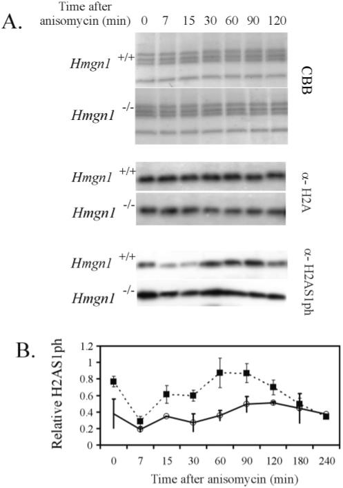
Loss of HMGN1 elevates the levels of H2A1ph throughout the anisomycin-induced stress response. (A) Extracts from Hmgn1+/+ and Hmgn1-/- mouse embryonic fibroblasts, obtained at various times after stimulation of quiescent cells with anisomycin, were analyzed with either anti-H2A or anti-H2AS1ph antibodies (indicated on the right of Western blot panels). (B) Quantitative analysis of the levels of H2A1ph after anisomycin treatment. X-ray films of developed Western blots were scanned and quantified, and the values of the H2A1ph signal were normalized to those of the corresponding H2A signals. The standard deviation values were calculated from four independent measurements.