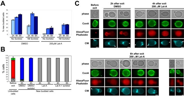
Figure 3. Bud emergence in the absence of F-actin.
Wild type cells were grown 7 days in YPDA medium at 30°C. Cells were incubated with Con-A-FITC for 1 h and then washed with “old” YPDA. Lat-A (200 µM final concentration) or DMSO were added and cells were further incubated at 30°C for 30 min. Cells were then re-fed with YPDA medium (with or without 1 M sorbitol) containing either 200 µM Lat-A or DMSO and grown at 30°C as described in material and methods. (A) Percentage of new budded cells after exit from quiescence at 30°C, N>200 for each time point, 2 experiments – error bars show SD. (B) Actin cytoskeleton organization in unbudded cells before exit from quiescence or in new budded cells after exit from quiescence at 30°C in YPDA medium containing either DMSO, DMSO and 1 M sorbitol, 200 µM Lat-A or 200 µM Lat-A and 1 M sorbitol. Red: Actin Bodies; green: depolarized actin cytoskeleton; blue: polarized actin cytoskeleton; grey: no detectable F-actin containing structures (N>200 for each time point, 2 experiments – error bars show SD). (C) Images of typical treated and untreated cells. The upper left panel display a typical wild type cell pre-incubated 30 min with Lat-A before re-feeding and the lower right panel, examples of Lat-A treated wild type cells 6 h after re-feeding with a collapsed new bud. Arrows indicate bud scar, arrowhead indicate birth scars; CW: Calcofluor White. Bar 2 µm. Of note, for AlexaFluor Phalloidin images, the maximum intensity is about 20 times lower for Lat-A treated cells than for untreated cells and was therefore greatly enhanced in the figure to document the absence of F-actin structure.

