
Figure 2:
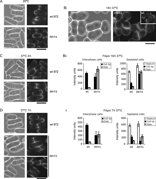
btn1Δ cells have altered sterol-rich membrane domains.Loss of polarization of sterol domains in btn1Δ cells at 37°C: filipin staining of wild-type 972 and btn1Δ cells grown at A) 25°C and Bi) 37°C for 18 h (left panel, phase contrast; inset, wild-type 972). Bii) Spatial distribution graphs of filipin density of indicated strains grown at 37°C for 18 h for interphase and septated cells, n = 25. Onset of filipin staining is linked to completion of cytokinesis: filipin staining of wild-type 972 and btn1Δ cells grown at 37°C for C) 4 h and Di) 7 h. Dii) Spatial distribution graphs of filipin density of indicated strains grown at 37°C for 7 h for interphase and septated cells, n = 25 individual measurements. Scale bar, 10 μm.