Figure 6:

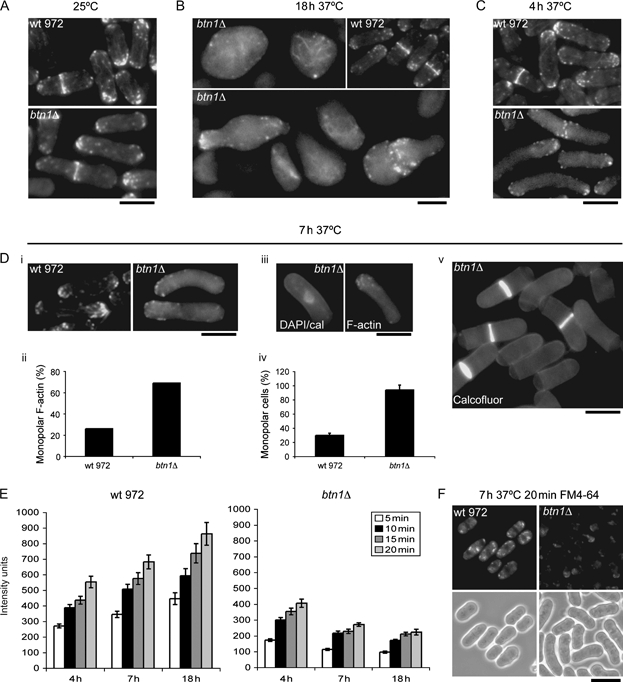
btn1Δ cells are defective in F-actin patch formation/polarisation and endocytosis. btn1 Δ cells have slight defects in the F-actin cytoskeleton at 25°C. A) Rhodamine–phalloidin actin staining of wild-type 972 and btn1Δ cells grown at 25°C. btn1 Δ cells develop severe defects in the F-actin cytoskeleton at 37°C. B) Loss of polarisation of F-actin patches in btn1Δ cells grown for 18 h at 37°C (inset, wild-type 972 grown under same conditions). C) Actin staining of wild-type and btn1Δ cells grown for 4 h at 37°C. Monopolar F-actin patch localization causes NETO defects in btn1Δ cells prior to total depolarized growth at 37°C. Di) Rhodamine–phalloidin actin staining of wild-type 972 and btn1Δ cells grown for 7 h at 37°C, ii) graph showing the percentage of indicated interphase cells with monopolar (rather than bipolar) actin (n = 300), iii) calcofluor/DAPI (left) and rhodamine–phalloidin F-actin (right) staining of btn1Δ cells grown for 7 h at 37°C, iv) graph showing the percentage of indicated cells monopolar for growth (by calcofluor staining) (three replicates of n = 300) and v) calcofluor staining of btn1Δ cells grown for 7 h at 37°C showing monopolar growth caps. btn1 Δ cells are severely defective for endocytosis at 37°C. E) Graph of FM4-64 uptake of wild-type and btn1Δ cells grown for indicated times at 37°C, displayed in intensity units over time. F) Panel showing 20 min FM4-64 uptake of indicated cells grown at 37°C for 7 h. Scale bar, 10 μm.
