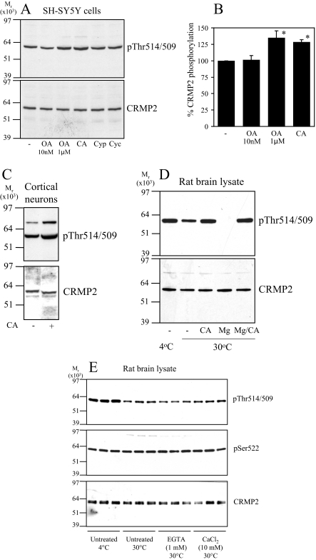
FIGURE 2.
Dephosphorylation of CRMP2 in cells. A, SH-SY5Y neuroblastoma cells were incubated with DMSO (control), okadaic acid (10 nm or 1 μm), calyculin A (CA, 10 nm), cypermethrin (Cyp, 10 μm), or cyclosporin A (Cyc, 10 μm) for 30 min. Cell lysates were subjected to Western blot analysis using Thr(P)-514/509 (upper panel) and total CRMP2 (lower panel) antibodies. The ratio of phospho-CRMP2/total CRMP2 in cells treated with okadaic acid or calyculin A compared with control cells is presented in B (n = 3; *, p < 0.05 relative to control (Students t test); average ± S.D.). C, primary rat cortical neurons were treated without or with calyculin A (10 nm), then cell lysates were immunoblotted as described in A. D, rat brain lysate was left on ice (lane 1) or incubated at 30 °C for 1 h (lanes 2-5) in the presence of 10 nm calyculin A (lane 3), 1 mm MgCl2 (lane 4), or 10 nm calyculin A plus 1 mm MgCl2 (lane 5). The lysates were immunoblotted as described in A. E, same as D, except lysates were incubated in the presence of 1 mm EGTA or 10 mm CaCl2 as shown.

