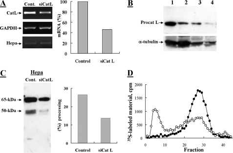
FIGURE 2.
Knockdown of procathepsin L in MDA-MB-435 cells inhibits processing of proheparanase. MDA-MB-435 cells were subjected to two sequential transfections with 2 μm anti-procathepsin L siRNA at a 48-h interval (siCat L), or were mock transfected and treated with reagents alone (Control). A, semi-quantitative RT-PCR analysis of cathepsin L (Cat L), heparanase (Hepa), and glyceraldehyde-3-phosphate dehydrogenase (GAPDH). Densitometric analysis (right) revealed a 54% decrease in the mRNA level of siCat L-treated cells versus control cells. Cells were lysed and subjected to Western blot analysis of: B, procathepsin L (lanes 1 (Control) and 2 (siCatL): 100 μg of cell lysate; lanes 3 (Control) and 4 (siCatL): 25 μg and cell lysate); and C, heparanase (pAb 1453), as described under “Materials and Methods.” Proheparanase processing, indicated by the generation of a 50-kDa subunit, was decreased (2.2-fold) in siCat L-transfected cells compared with mock transfected cells, as revealed by densitometry analysis (right panel). D, heparanase activity. siCat L (○) and mock (•) transfected cell lysates were incubated with 35S-labeled ECM and analyzed for heparanase enzymatic activity, as described in the legend to Fig. 1.

