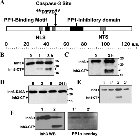
FIGURE 1.
Inhibitor-3 is cleaved by caspase-3 in vitro at a DTVD cleavage site. A, domain map of human Inh3. The diagram shows the location of the putative caspase-3 cleavage site (46DTVD49), the nuclear localization signal (NLS), the nucleolar targeting signal (NTS) (19), the PP1 binding motif (39KKVEW43) (18), and the inhibitory domain that lies between residues 64 and 77. B, in vitro cleavage of Inh3 by caspase-3. Purified recombinant His6-Inh3 was incubated with recombinant human caspase-3 for the indicated times and Western blotted with a rabbit polyclonal antibody against Inh3 (see “Experimental Procedures”). Inh3-CT is the 17.5-kDa cleavage product. C, purified recombinant Inh3 was treated with caspase-3 as in B, except that a peptide-specific antibody to amino acids 69–83 of Inh3 was used. D, mutation of the caspase-3 site in Inh3 renders it resistant to cleavage. Recombinant Inh3(D49A) was treated with caspase-3 as in B, and the digests were analyzed by Western blotting with a polyclonal antibody against Inh3. One representative experiment of three is shown in A–D. E, the Inh3-CT cleavage product does not immunoprecipitate with PP1. Purified Inh3 or Inh3 predigested with caspase-3 was incubated with PP1α for 1 h at 4 °C, immunoprecipitated with PP1α antibody, and Western blotted with anti-Inh3 antibody (see “Experimental Procedures”). Lane 1, Inh3 input; lane 1′, immunoprecipitate of PP1α plus Inh3; lane 2, caspase-3-cleaved Inh3 input; lane 2′, immunoprecipitate of PP1α plus caspase-3-cleaved Inh3. F, overlay assay. Left, Western blot of Inh3 and caspase-3-treated Inh3 with Inh3 antibody. Lane 1, Inh3; lane 2, caspase-3-cleaved Inh3. Right, overlay blot of Inh3 and caspase-3-treated Inh3 with PP1. Lanes 1′ and 2′ correspond to lanes 1 and 2 in the left panel. The membranes were blocked with 5% nonfat milk proteins and then incubated with purified PP1α. The membrane was washed and then probed with anti-PP1 antibody (see “Experimental Procedures”). Essentially identical results were obtained in two independent experiments for the data shown in E and F. a.a., amino acids; WB, Western blot.

