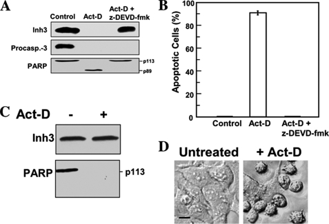
FIGURE 3.
Inh3 is not degraded during act-D-induced apoptosis in the presence of the caspase inhibitor, Z-DEVD-fmk, or in a caspase-3-deficient cell line. A, HL-60 cells were incubated with or without caspase inhibitor (Z-DEVD-fmk; 100 μm) for 1 h and then treated with 4 μm act-D for an additional 5 h to induce apoptosis. Cell lysates were subjected to Western blot analysis using antibodies against Inh3, caspase-3, and PARP. B, the percentage of apoptotic cells was determined by the Hoechst assay (see “Experimental Procedures”). The error bar indicates the mean ± S.D. (n = 3). C, MCF-7 cells, which lack caspase-3, were treated with 16 μm act-D for 24 h. Rounded and detached cells were collected, lysed, and Western blotted for Inh3 and PARP. D, phase-contrast microscopy of MCF-7 cells treated as in C, showing the morphological differences between untreated and act-D-treated cells. Phase images were obtained using a Zeiss Axioplan 2 fluorescence microscope with a ×20 objective (see “Experimental Procedures”). Scale bar (black horizontal bar in the left image), 10 μm.

