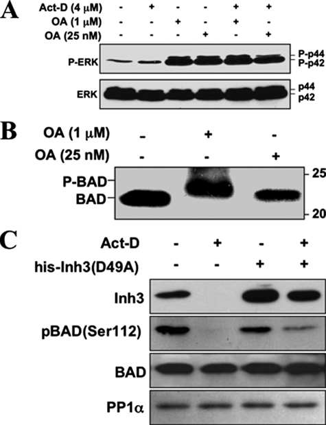
FIGURE 7.
PP1 activity is involved in the dephosphorylation of BAD, and BAD dephosphorylation during act-D-induced apoptosis is inhibited by electroporation of Inh3(D49A). A, HL-60 cells were treated with 1 μm or 25 nm OA for 5 h, in the absence and presence of act-D (4 μm), as indicated. Cell lysates were Western blotted with a phosphospecific antibody for ERK (top), and with an antibody against ERK (bottom). B, HL-60 cells were treated with 1 μm or 25 nm OA for 5 h. Cells were then analyzed for BAD by Western blotting. The positions of phosphorylated (P-BAD) and nonphosphorylated BAD are indicated. Data for A and B are representative of three experiments. C, HL-60 cells were electroporated with or without purified His-Inh3(D49A) prior to treatment with act-D for 5 h or without act-D, as indicated. Cell lysates were Western blotted for Inh3, BAD-phosphoserine 112, BAD, and PP1α. The data are representative of three independent experiments.

