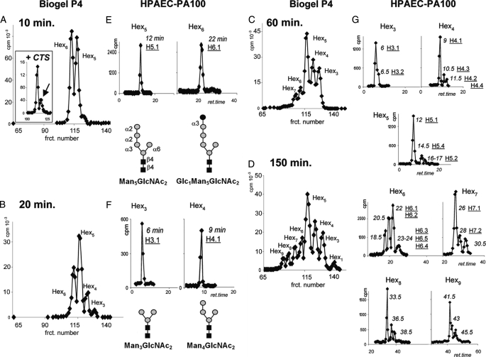
FIGURE 2.
The complexity of N-glycans made by E. histolytica trophozoites increases dramatically with time. E. histolytica trophozoites were incubated with [2-3H]Man for 10, 20, 60, and 150 min, and N-glycans were released with PNGase F and separated by Bio-Gel P-4 filtration (A-D), where Hexn indicates their size. Isomers with the same number of hexoses were further isolated on an HPAEC-PA100 column (E-H), with the retention time (ret.; min) shown in italic and the identifier name from Fig. 1 (e.g. H5.1) underlined below. After 10-min labeling (A and E), the predominant E. histolytica N-glycans were unmodified Man5GlcNAc2 and the glucosylated product of UDP-Glc:glycoprotein glucosyltransferase (Glc1Man5GlcNAc2) (H6.1). As shown in the inset in A, Glc1Man5GlcNAc2 is by far the most abundant N-glycan in the presence of the glucosidase II inhibitor castanospermine (CTS). After 20-min labeling (B and F), two mannosidase digestion products are apparent: Man4GlcNAc2 (H4.1) and Man3GlcNAc2 (H3.1). After 60-min labeling (C), complex N-glycans are apparent in Hex6 and Hex7 pools. After 150-min labeling (D and G), novel, complex N-glycans are present in all of the pools (see further characterization in Fig. 4). frct., fraction.

