
Fig. 1.
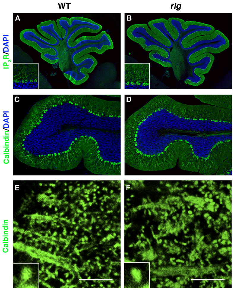
Immunohistochemical staining for Purkinje cell (PC) specific markers in the cerebellum of rig mice. (A, B) Wild-type (A) and rig (B) cerebellum stained with anti IP3 receptor (IP3R, green). Insets show higher magnification view of PCs. (C, D) The lobule IX of wild-type (C) and rig (D) cerebellum stained with anti Calbindin-D (green). Cell nuclei are counterstained with DAPI (blue) in A–D. (E, F) Calbindin-D staining showing the morphology of PC dendritic spines in wild-type (E) and rig (F) mice. Insets show higher magnification view of individual spines. Scale bars: 5 μm.