
Fig. 3.
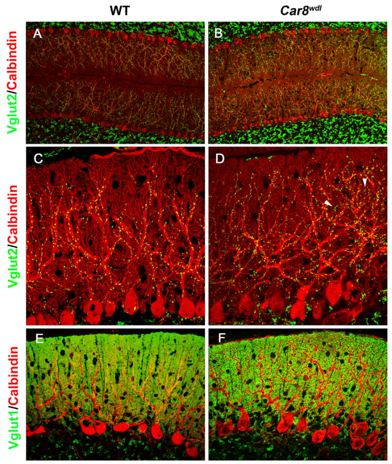
CF and PF labeling in the wild-type (A, C, E) and Car8wdl (B, D, F) cerebellum. (A, B) Double immunofluorescence for VGLUT2 (green) and Calbindin (red). Note that the distribution of VGLUT2-labeled CF terminals is expanded distally in the molecular layer of Car8wdl (B) compared to wild-type (A) mice. (C, D) Higher magnification view of VGLUT2 (green) and Calbindin (red) labeling. While VGLUT2 is mainly localized along the thick, proximal dendrites in wild-type mice (C), VGLUT2-labeled terminals are also associated with thinner, distal dendrites in Car8wdl mice (D, arrowheads). Note an increase of VGLUT2-labeled puncta in the Car8wdl molecular layer. (E, F) Double imunofluorescence for VGLUT1 (green) and Calbindin (red). No overt difference in the distribution of VGLUT1-labeled PF terminals was detected between wild-type (E) and Car8wdl (F) mice.