
Fig. 5.
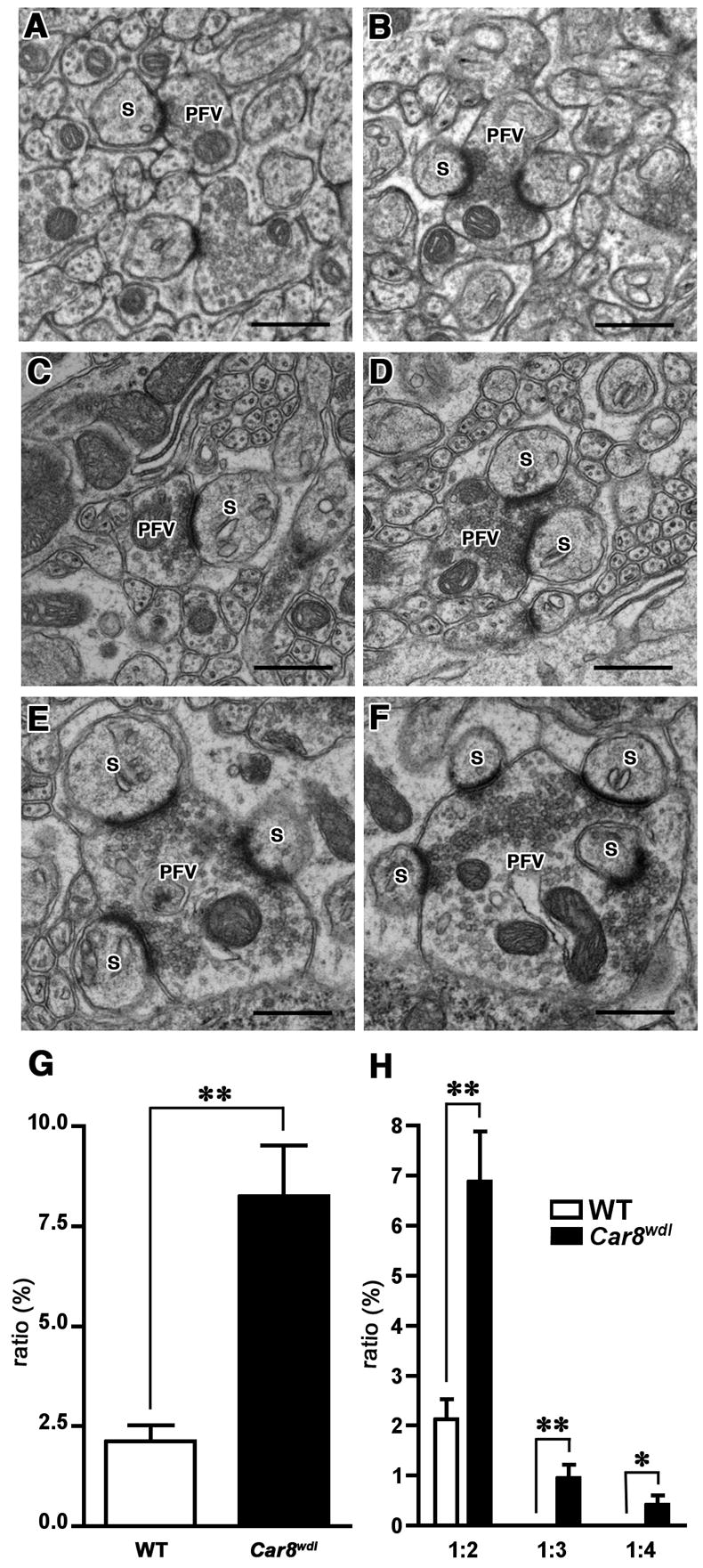
Electron microscopic analysis of excitatory synaptic contacts in wild-type and Car8wdl cerebellum. In wild-type mice, parallel fiber varicosities interact with one (A) or two (B) dendritic spines of the PCs. In the Car8wdl cerebellum, in addition to the varicosities interacting with one (C) or two (D) dendritic spines, those contacting more than two spines (E, F) are observed. S: dendritic spines, PFV: parallel fiber varicosity. Scale bar: 500 nm. As summarized in (G), the percentage of multiple synaptic varicosities (2–4 dendritic spines contacting 1 varicosity) against total synaptic interactions is significantly higher (**, p<0.005) in Car8wdl mice. (H) shows the classification of multiple synaptic varicosities into 1:2 (presynapse vs postsynapse), 1:3 and 1:4, and the percentage of multiple synaptic varicosities belonging to each class. ** and * denote statistical significance with p<0.005 and p<0.05, respectively.