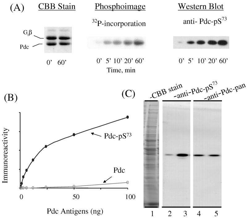
Fig. 1. Characterization of antibody AN519 (anti-Pdc-Ser73).
A. Time course of purified bovine Pdc phosphorylation by PKA, analyzed by autoradiography and Western Blotting. B. Specificity of AN519 for phosphorylated bovine Pdc. Increasing amounts of phosphorylated (Pdc-pSer73) or non-phosphorylated (Pdc) bovine phosducin were subjected to Western blot analysis with AN519. C. Phosphorylation of Pdc in mouse retinal homogenate. Mouse retinal extract (40 μg) was incubated with ATP/Mg, okadaic acid, IBMX with (lanes 1, 3, 5) or without cAMP (lanes 2, 4) followed by SDS/PAGE. The proteins were visualized either by Coomassie brilliant blue (CBB) staining or Western blot analysis with AN519 or anti-Pdc-pan. See Materials and methods for details.

