
Figure 1.
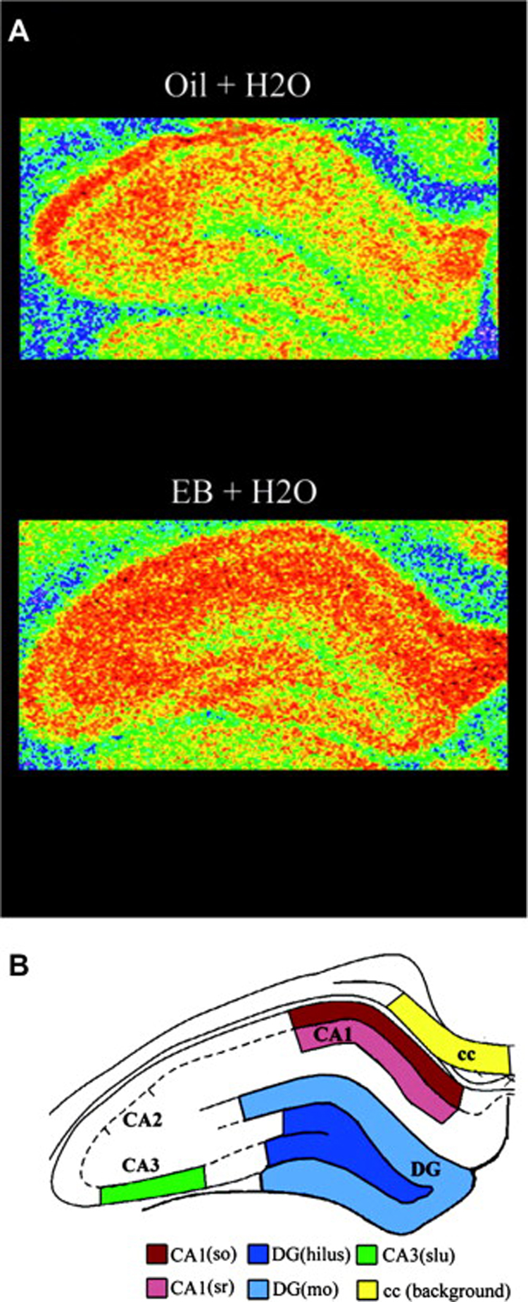
Estradiol increases spinophilin expression in the hippocampal formation of ovariectomized female rats. A, Illustration of autoradiograms using pseudocolor representation of shading densities of spinophilin immunoreactivity in the hippocampal formation of representative estrogen (EB)- and control (Oil)-treated ovariectomized rats (blue < green < yellow < orange). B, Schematic diagram identifying specific hippocampal regions from where measures were taken. cc, corpus callosum; dg, dentate gyrus; (so), stratum oriens; (sr), stratum radiatum; (slu); stratum lucidum; (mo), molecular layer. Reprinted with permission from Brake et al., 2001.