
Figure 3.
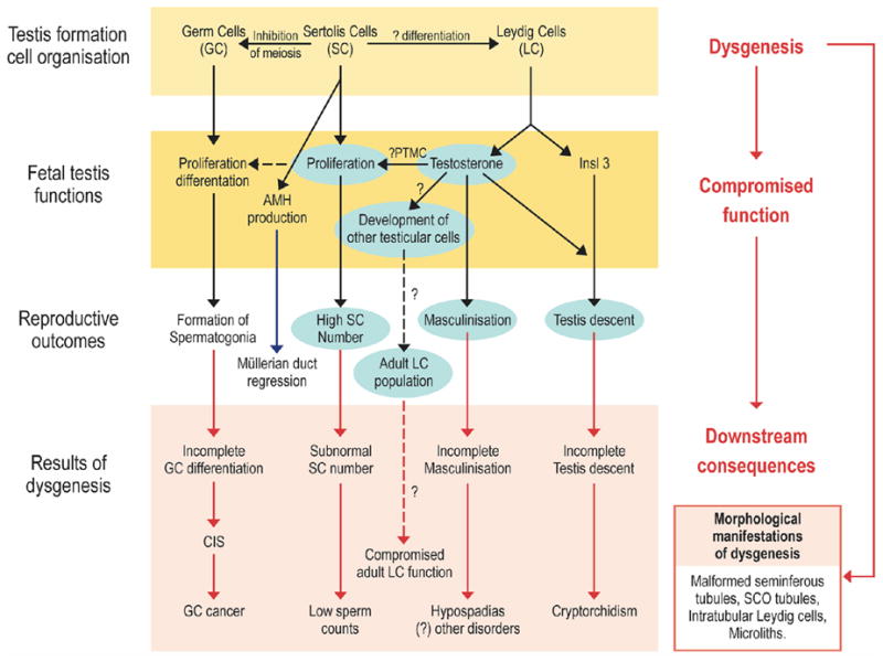
Schematic diagram to illustrate how dysgenesis of the early fetal testis is thought to lead to abnormalities of somatic cell function, resulting in hormonal changes and the downstream disorders that comprise testicular dysgenesis syndrome (TDS). The central role of testosterone is highlighted by the blue boxes. Dashed lines show pathways that are hypothesized but unproven (68).
Abbreviations: PTMC=peritubular myoid cell; Insl3=insulin-like factor 3; AMH=anti-mullerian hormone; CIS=carcinoma-in-situ; SCO=Sertoli cell only.