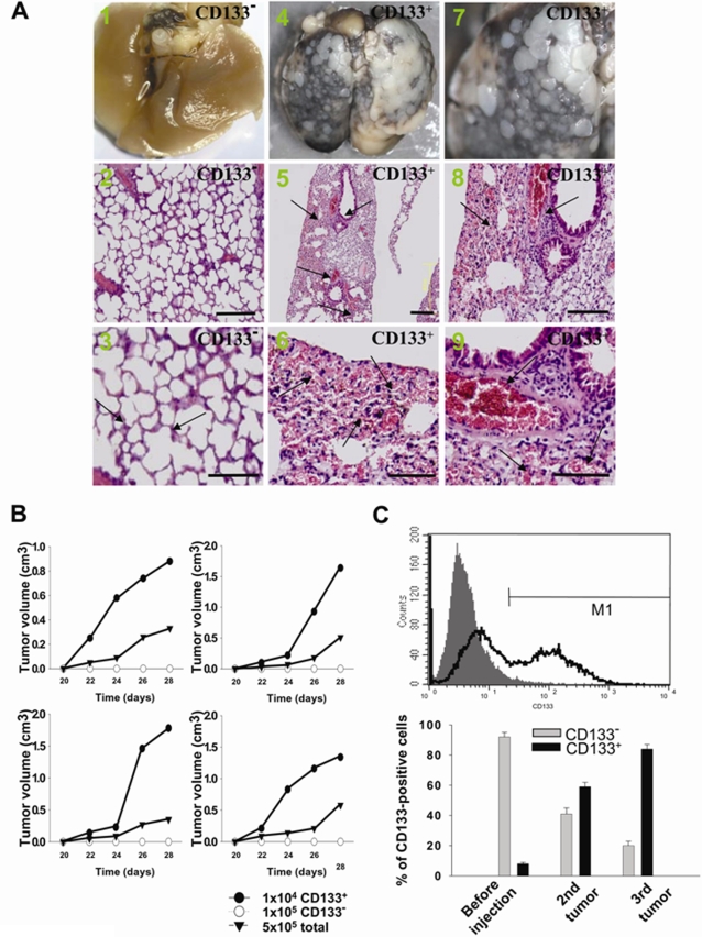
Figure 3. Evaluation of the tumorigenicity of LC-CD133+ and LC-CD133− in vivo.
(A) The in vivo tumorigenicity of LC-CD133+ and LC-CD133− in tail vein-injected mice was analyzed by macroscopic and histological examination. A1–3: LC-CD133−; arrows: normal alveolar structure of lung. A4–6: LC-CD133+; arrows: tumor formation. A7–9: LC-CD133+; arrows: neovascularity and thrombosis. Bar: 200 µm. (B) The in vivo tumor-restoration and proliferative ability of 104 LC-CD133+, 105 LC-CD133− and 5×10⁵ total tumor cells from patient No. 1, 2, 4, and 7 were examined by xenotransplanted tumorigenicity analysis. (C) The tumor repopulation ability of LC-CD133+ was studied in transplanted SCID mice. The expression levels of CD133 were determined by FACS analysis from primary LC-CD133+, second tumor, and third tumor. Data shown here are the mean±SD of three experiments.

