
Figure 2.
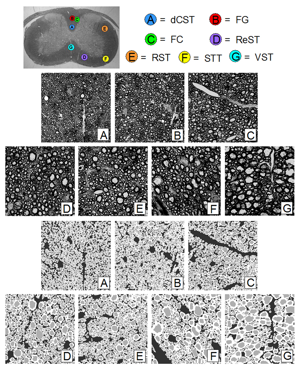
Top Image: Optical image of C6/C7 mouse cord section showing WM tract locations: A) dorsal corticospinal (dCST), B) gracilis (FG), C) cuneatus (FC), D) rubrospinal (RST), E) spinothalamic (STT), F) reticulospinal (ReST), G) vestibulospinal (VST). The spinal cord is approximately 3 mm wide. Middle Images: Optical images of WM tracts from mouse spinal cord C6/C7 section. Each image is 700 × 700 with a pixel resolution of 0.1 × 0.1 µm. Bottom Images: Segmented down-sampled images of WM tracts from mouse spinal cord C6/C7 section. Each image is 256 × 256 with a pixel resolution of 0.27 × 0.27 µm.