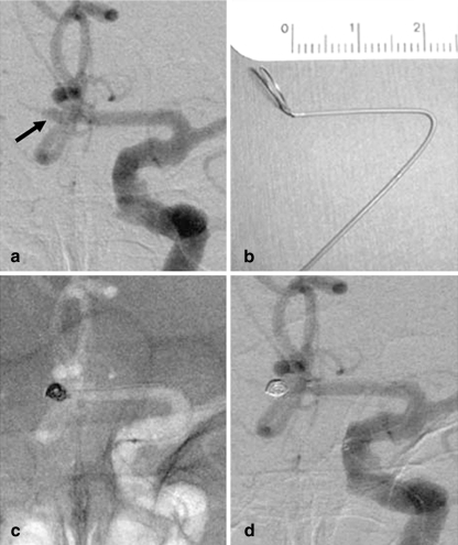
Fig. 1.
Patient 4. a Angiogram of the left ICA shows a tiny ACoA aneurysm with a maximum diameter of 2.5 mm (arrow). b Photograph of a steam-shaped microcatheter. The shaping mandrel is bent to conform to the shape of the horizontal portion of the anterior cerebral artery. c Unsubtracted image of the skull obtained with the road-mapping technique during treatment. Embolization was with a GDC-10 Soft coil measuring 2×60 mm. d Angiogram of the left ICA obtained at the end of the procedure shows complete occlusion of the aneurysm. The diameter of the coil mass was larger than that of the aneurysm sac before embolization, indicating aneurysm distention

