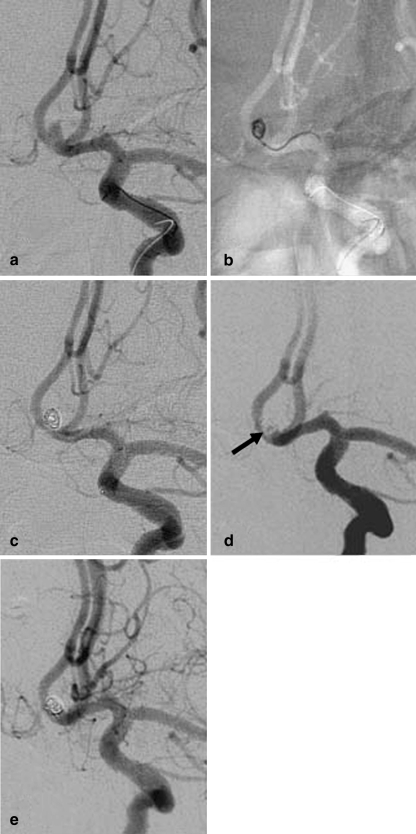
Fig. 2.
Patient 5. a Angiogram of the left ICA shows a tiny ACoA aneurysm with a maximum diameter of 2.5 mm. b Unsubtracted image of the skull obtained with the road-mapping technique during treatment. Embolization was with a GDC-10 Soft coil measuring 2×60 mm. c Angiogram of the left ICA obtained at the end of the procedure shows complete occlusion of the aneurysm. d Angiogram of the left ICA obtained at the 3-month follow-up shows minor recanalization of the neck (arrow). e The minor neck filling was stable at the 48-month follow-up

