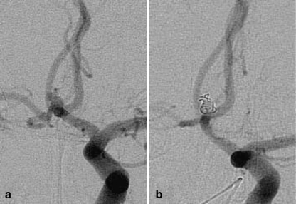
Fig. 3.
Patient 8. a Angiogram of the left ICA shows a tiny ACoA aneurysm with blebs. b Angiogram of the left ICA obtained at the end of the procedure. The aneurysm sac and blebs are completely occluded with coils. The coil mass is larger than the aneurysm sac before embolization (indicating aneurysm distention)

