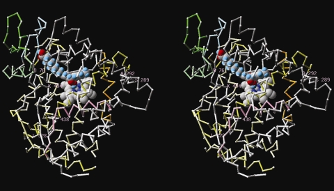
Fig. 6.
3-D-structure of the StAOS2 protein in complex with heme and the substrate molecule. Steroscopic view of the predicted spatial localization of residues conserved within QR alleles StAOS2-1 and StAOS2-6, which differ in QS alleles StAOS2-7 and StAOS2-8. heme and the 13-(S)HPOTE (13-hydroperoxyoctadecatrienoic acid) substrate molecule placed manually based on the position occupied by the palmitoyl-glycine molecule present in Bacillus megaterium CYP102 structure (1jpza) appear in spacefilling. All positions are indicative only. Numbering is according to StAOS2-1; color scheme according to Table S2

