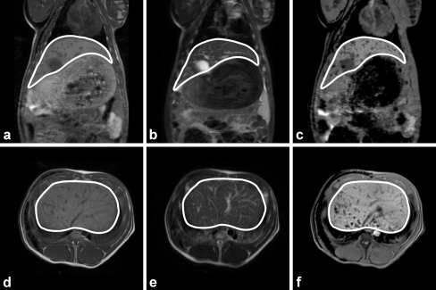
Fig. 6.
In vivo coronal (a–c) and transverse (d–f) MR images of pig 17. The anatomical images are found in a and d (T 1-weighted SE), and b and e (T 2-weighted SE images). c and f The holmium-sensitive T 2*-weighted GE images. In the T 1-weighted SE images, the vessels and gallbladder show up as hypo-intense structures and as hyperintense structures in the T 2-weighted SE images. In the T 2*-weighted GE images, the clusters of microspheres are visualized as additional focal regions of signal loss, especially in the dorsal region

