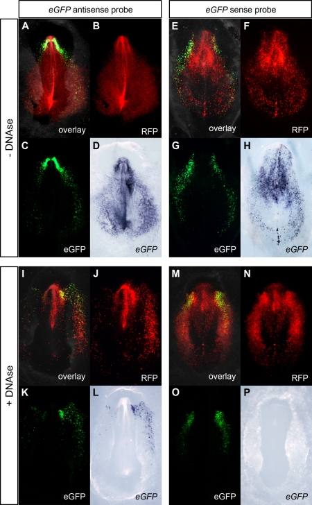
Figure 2. Comparison between eGFP fluorescence and eGFP expression patterns in Cer0.4-eGFP/CAGGS-RFP electroporated chick embryos.
Chick embryos were co-electroporated with Cer0.4-eGFP (tissue-specific reporter) and pCAGGS-RFP (ubiquitous reporter) at stage HH3 and processed for WISH. (A, E, I, M) Merge of bright field with fluorescence images. (B, F, J, N) RFP fluorescence. (C, G, K, O) eGFP fluorescence. (D, H, L, P) WISH using eGFP antisense and sense probes. At stages HH6-7, RFP fluorescence was detected in all electroporated cells, whereas eGFP fluorescence was specifically observed in the anterior mesendoderm. When embryos were processed by the standard WISH method, the eGFP antisense probe was detected in both eGFP- and RFP-positive cells (D). These cells were also labeled by the eGFP sense probe (H). In embryos treated with DNase I, the antisense probe was restricted to the eGFP-expressing cell population (L), whereas the sense probe was no longer detected (P).

