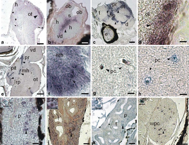
Fig. 1.
Expression of a glucocorticoid receptor (DlGR1) in tissues of larval stages of sea bass (Dicentrarchus labrax). Horizontal sections (unless otherwise indicated). a–g, i, j, l In situ hybridization (ISH; black arrowheads) with DlGR1 riboprobe. h, k Immunohistochemistry (IH; black arrowheads) with anti-DlGR1 specific antibody. a Pre-larva at 3 dph, optic lobe, ISH (ot optic tectum, h @brain hemisphere). Bar 100 μm. b Larva at 10 dph, brain, ISH (h brain hemisphere, vd area ventralis telencephali pars dorsalis, s scissure, ob olfactory bulb). Bar 100 μm. c Post-larva at 10 dph, sagittal section of brain, ISH (t telencephalon, pr preoptic region, np nucleus preopticus, d diencephalon, e eye). Bar 100 μm. d Post-larva at 30 dph, optic lobe, ISH (ot optic tectum). Bar 20 μm. e Post-larva at 50 dph, sagittal section of brain, ISH (pit pituitary gland, pr preoptic region, mh mediobasal hypothalamus, ot optic tectum, vd area dorsalis telencephali pars dorsalis, rp posterior recess). Bar 250 μm. f Juvenile at 50 dph, detail of the mediobasal hypothalamus (mh), ISH. Bar 20 μm. g Juvenile at 50 dhp, brain transverse section, ISH (pc pyramidal cell). Bar 20 μm. h Juvenile at 50 dph, brain transverse section, IH (pc pyramidal cell). Bar 20 μm. i Larva at 25 dph, head kidney, ISH (p parenchyma). Bar 40 μm. Inset: Parenchymatic cell (pc). Bar 20 μm. j Juvenile at 50 dph, head kidney, ISH (p parenchyma). Bar 80 μm. Inset: Parenchymatic cell (pc). k Juvenile at 50 dph, head kidney, IH (p parenchyma, t tubule). Bar 80 μm. Inset: Parenchymatic cell (pc). Bar 25 μm. l Post-larva at 36 dph, spleen, ISH (wpc white pulp cells). Bar 60 μm. Inset: White pulp cell (wpc). Bar 20 μm

