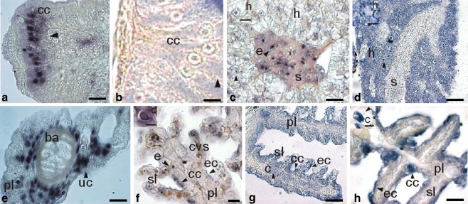
Fig. 2.
Expression of a GR (DlGR1) in tissues of larval stages of sea bass (D. labrax). Horizontal sections (unless otherwise indicated). a, c, e, f In situ hybridization (ISH; black arrowheads) with DlGR1 riboprobe. b, d, g, h Immunohistochemistry (IH; black arrowheads) with anti-DlGR1 specific antibody. a Juvenile at 50 dph, anterior intestine, ISH (cc mucosa columnar cells). Bar 60 μm. b Juvenile at 50 dph, anterior intestine, IH (cc mucosa columnar cells). Bar 20 μm c Juvenile at 50 dph, liver, ISH (s sinusoid, h hepatocyte, e erythrocyte). Bar 40 μm. Inset: Hepatocyte (h). Bar 20 μm. d Juvenile at 50 dph, liver, IH (h hepatocyte, s sinusoid). Bar 100 μm. Inset: Hepatocyte (h). Bar 20 μm. e Larva at 12 dph, gills, ISH (uc undifferentiated cells, ba branchial arch, pl primary lamellae). Bar 60 μm. f Post-larva at 36 dph, gills, ISH (cvs central venous sinus, sl secondary lamellae, ec epithelial cells, cc chloride cells, e erythrocytes, pl primary lamellae). Bar 40 μm. Inset: Chondrocyte (c). Bar 20 μm. g Larva at 20 dph, gills, IH (c chondrocyte, ec epithelial cells, cc chloride cells, sl secondary lamellae, pl primary lamellae). Bar 40 μm. h Post-larva at 36 dph, gills, IH (cc chloride cells, ec epithelial cells, pl primary lamellae, sl secondary lamellae). Bar 40 μm. Inset: Chondrocyte (c). Bar 20 μm

