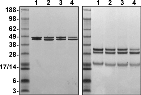
FIGURE 1.
SDS-PAGE analysis. Purified proteins (4 μg/lane) were subjected to SDS-PAGE under non-reducing (left) or reducing (right) conditions and visualized by staining with Coomassie Blue R-250. Lane 1, human PDFXa; lane 2, rFXa; lane 3, rFXaV17A; and lane 4, rFXaI16L. The apparent molecular weights of the standards are indicated on the gels.

