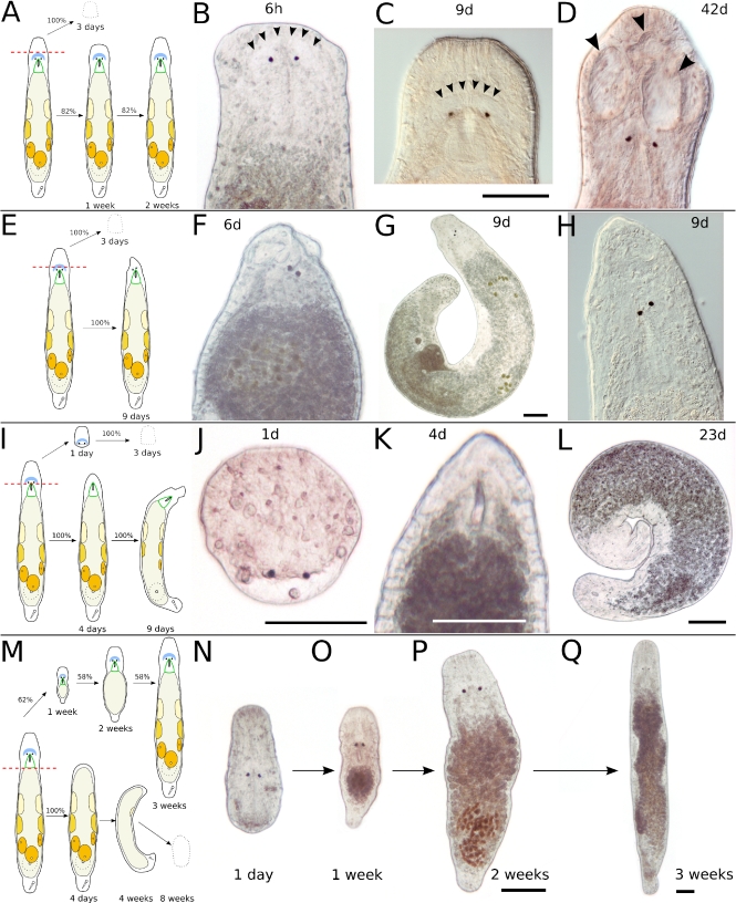
Fig. 3.
Transversal amputation in the rostrum (a–d), in the brain (e–h), behind the eyes (i–l), and at the end of the pharynx (m–q). a Schematic drawing of amputation in the rostrum. The anterior piece perishes within about 3 days; the posterior piece regenerates in about 2 weeks. b Six hours after amputation, the brain is intact. Arrowheads mark the anterior border of the neuropil. c Same specimen as in b, 9 days after amputation. The rostrum is not yet at full length. Arrowheads mark the anterior border of the neuropil. d Different specimen, 42 days after amputation. The rostrum is at full length but developed grooves (arrowheads). e Schematic drawing of amputation in the brain. The anterior piece perishes in about 3 days; the posterior piece fails a complete regeneration. f Six days after amputation. g Different specimen, 9 days after amputation. h Detail of specimen in g. Note loose rostrum and skewed eyes (f–h). i Schematic drawing of amputation directly behind the eyes. The anterior piece perishes in about 3 days; the posterior piece does not regenerate eyes and a proper rostrum. j Anterior piece 1 day after amputation. The wound is closed, but no blastema is formed. The space behind the eyes is acellular and fluid-filled. k Posterior piece of same specimen 4 days after amputation. Note the blastema-like tissue anterior of the pharynx, which does not regenerate eyes and brain. l Same specimen, 23 days after amputation. The animal is curled up, with an incompletely regenerated rostrum. Gonads are still present. m Schematic drawing of amputation at the end of the pharynx. The anterior piece regenerates to a complete animal in 58% of cases. n–q Same specimen at different time intervals after amputation. All scale bars are 100 μm. Same scale bar in b–d, f, h, and n–p

