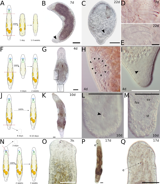
Fig. 4.
Transversal amputation in or behind the pharynx and at the tail plate (a–e), between testes and ovaries (f–i), and at the tail plate (h–m). Oblique amputation between the eyes (n–q). a Schematic drawing of amputation in or behind the pharynx and at the tail plate. Only the centerpiece is observed, which will regenerate a tail plate with duo-gland adhesive systems and often also a stylet, in the absence of a brain. b Seven days after amputation, a tail plate with developing stylet (arrowhead) has regenerated. c Another specimen, 22 days after amputation. A tail plate and a stylet (arrowhead) have regenerated, but the stylet is small and misplaced. d Stylet, detail of b. e Stylet, detail of c. f Schematic drawing of amputation between testes and ovaries. Only the anterior piece is observed. After 4 days, a small stylet has appeared in the tail plate, and the testes resume spermatogenesis. g Four days after amputation. h Left testis, detail of g. Note the round spermatids in the center of the testis (arrowheads). i Tail plate, detail of g. Arrowhead points at small stylet. j Schematic drawing of amputation at the tail plate. The posterior piece will die within 8 days; the anterior piece will regenerate within 6–10 days. k Fully regenerated animal 10 days after amputation. l Detail of the tail plate of another specimen after 10 days of regeneration. About 140 duo-gland adhesive systems at the rim of the tail plate. Arrowhead points at male genital opening. m Same animal as in l, different focal plane. fsv false seminal vesicle, st stylet, sv seminal vesicle. n Schematic drawing of oblique amputation between the eyes. The anterior piece perishes in about 3 days; the posterior piece does not regenerate the missing eye. o Amputated specimen 7 h after amputation. p Same specimen as in o, 17 days after amputation. Note that the gonads are fully developed at the side of the missing eye. q Detail of p. All scale bars are 100 μm, except 10 μm in e. Sub-panels d–e and h–i are of the same scale

