
Fig. 10. Protection of ERK activity by pORF3.
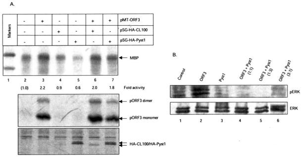
A, cells were transfected with the indicated plasmids, and the endogenous ERK activity present in cell lysates was determined using the MBP assay. The -fold activity was calculated by densitometric scanning of the bands; the ERK activity present in mock-transfected cells (lane 2) was used as a reference. The lower panels show Western blots for expression of pORF3 and HA-tagged CL100 or Pyst1. Arrows indicate the relevant protein bands. B, cell lysates were Western blotted for cellular ERK with anti-phospho-ERK (pERK) or anti-ERK antibodies.