
Fig. 2. Effect of various pathway inhibitors on pORF3-mediated ERK activation.
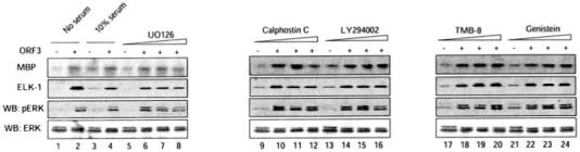
Cells stably expressing pORF3 or a control cell line were serum-starved and then treated with either 10% serum (lanes 3 and 4) or with one of the inhibitors indicated (lanes 5-24). Cell growth and treatments were as described under “Experimental Procedures.” For each set, the control cells were treated with an IC50 concentration of the inhibitor; the pORF3-expressing cells were treated with IC50, 2× IC50, or 5× IC50 concentrations of the inhibitor. Cell lysates were tested for ERK activity using MBP and Elk-1 as substrates as described under “Experimental Procedures.” The lysates were also Western blotted (WB) with anti-phospho-ERK and total ERK antibodies. Each blot was individually scanned, and the bands were quantitated by densitometry using Kodak 1D image analysis software (Kodak Digital Science).