
Fig. 8. ERK activation by mutant ORF3 proteins.
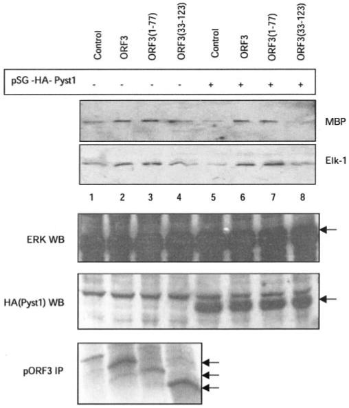
Cells were transfected with expression vectors for wild type (lanes 2 and 6) or mutant (lanes 3, 4, 7, and 8) ORF3 proteins in the absence (lanes 1-4) or presence (lanes 5-8) of plasmid pSG-HA-Pyst1. Empty pMT3 vector was used as control (lanes 1 and 5). Cell lysates were prepared and assayed for ERK activity using either MBP or Elk-1 as a substrate as described under “Experimental Procedures.” The lower panels show Western blots (WB) for expression of ERK and Pyst1 (anti-HA tag) and immunoprecipitation (IP) for full-length and deleted pORF3. Arrows indicate the relevant protein bands.