Figure 4.
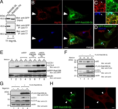
Effect of the constitutive active mutant of Rab33B on autophagy. (A) The constitutive active mutant Rab33B interacted with Atg16L. Beads coupled with T7-Atg16L were incubated with COS-7 cell lysates containing either GFP-Rab33B-QL + 0.5 mM GTPγS (lane 1) or GFP-Rab33B-TN + 1 mM GDP (lane 2). The lysates (top, input) and proteins bound to the beads (middle, IP) were analyzed by SDS-PAGE followed by immunoblotting with anti-GFP antibody (Blot: anti-GFP, top and middle panels) and HRP-conjugated anti-T7 tag antibody (Blot: anti-T7, bottom). Input means 1/80 volume of the reaction mixture used for immunoprecipitation (input, top). (B) GFP-Rab33B-QL induced Atg16L-positive punctate structures in the cytoplasm. NIH3T3 cells transiently expressing GFP-Rab33B-QL (green, large arrowheads) were stained with the anti-Atg16L antibody (red) and anti-GM130 antibody (blue). Bar, 10 μm. (C) High magnification of the boxed area in B. Colocalization of Rab33B-QL and Atg16L was evident in the punctate structures in the cytoplasm, but not in the Golgi. The arrows point to the position of the Golgi. (D) NIH3T3 cells transiently expressing GFP-Rab33B-TN (green) were stained with the anti-GM130 antibody (blue) and anti-Atg12 antibody (red). In contrast to the GFP-Rab33B-QL in (B), GFP-Rab33B-TN was exclusively localized in the Golgi (arrows). Bar, 10 μm. (E) Induction of LC3-lipidation by mRFP-Rab33B-QL. Cell lysates from HEK293 cells transiently expressing nothing (lanes 1 and 2), mRFP (lanes 3 and 4), mRFP-Rab33B-TN (lanes 5 and 6), or mRFP-Rab33B-QL (lanes 7 and 8) were analyzed by SDS-PAGE followed by immunoblotting with anti-LC3 antibody (Blot: anti-LC3, top panel), anti-mRFP antibody (Blot: anti-mRFP, middle panel), and anti-α-tubulin antibody (Blot: anti-tubulin, bottom panel). (F) Accumulation of p62 in FLAG-Rab33B-QL-expressing cells. Lysates from NIH3T3 cells (lanes 1 and 2) and NIH3T3 cells constitutively expressing FLAG-Rab33B-QL (lanes 3 and 4) under nutrient-rich conditions (lanes 1 and 3, medium “D”) or starvation conditions (lanes 2 and 4, medium “H”) were analyzed by SDS-PAGE followed by immunoblotting with anti-p62 antibody (Blot: anti-p62, top panel), anti-FLAG tag antibody (Blot: anti-FLAG, second panel from the top), anti-LC3 antibody (Blot: anti-LC3, third panel form the top), and anti-actin antibody (Blot: anti-actin, bottom panel). (G) Effect of nigericin. Lysates from NIH3T3 cells (lanes 1 and 2) and NIH3T3 cells constitutively expressing FLAG-Rab33B-QL (lanes 3 and 4) under nutrient-rich conditions in the presence of (lanes 2 and 4) or in the absence of (lanes 1 and 3) 20 μg/ml nigericin were analyzed by SDS-PAGE followed by immunoblotting with anti-FLAG tag antibody (Blot: anti-FLAG, top), anti-LC3 antibody (Blot: anti-LC3, middle), and anti-actin antibody (Blot: anti-actin, bottom). (H) GFP-Rab33B-QL did not induce LC3-positive punctate structures in the cytoplasm under nutrient-rich conditions. NIH3T3 cells transiently expressing GFP-Rab33B-QL (green, arrowheads) were stained with the anti-LC3 antibody (red). Bar, 10 μm.

