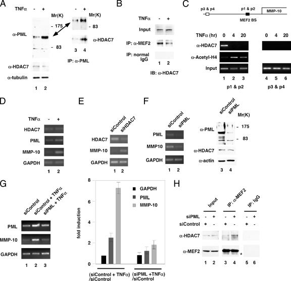
Figure 3.
TNF-α induces dissociation of HDAC7 from MEF2 and association of HDAC7 and PML. (A) HUVECs were treated with or without TNF-α, and whole cell extracts were prepared followed by immunoprecipitation with anti-PML antibodies and Western blotting with the indicated antibodies. Whole cell extracts (lanes 1 and 2) were immunoprecipitated with anti-PML antibodies and probed with anti-PML or anti-HDAC7 antibodies (lanes 3 and 4). One major band migrating at 110–120 kDa and one minor band migrating at 80 kDa were detected. (B) HUVEC whole cell extracts treated with TNF-α were immunoprecipitated with anti-MEF2 antibodies and probed with anti-HDAC7 antibodies. (C) Chromatin immunoprecipitation assays were carried out using anti-HDAC7 and anti-acetyl-histone H4 antibodies as described in Materials and Methods. Lanes 1–3, PCR using primers flanking the MEF2 binding sites (BS). Lanes 4 and 5, PCR using primers 2 kb upstream of the MEF2 BS. (D) HUVECs were treated with or without TNF-α as described in A, and total RNA was isolated followed by RT-PCR using gene-specific primers as indicated. (E) siRNA-mediated knockdown of HDAC7 was performed, and total RNA was isolated followed by RT-PCR using gene-specific primers as indicated. (F) siRNA-mediated knockdown of PML was performed, and total RNA was isolated followed by RT-PCR using gene-specific primers as indicated. (G) siRNA-mediated knockdown of PML was performed followed by TNF-α treatment and total RNA was isolated followed by RT-PCR using gene-specific primers as indicated. Real-time PCR was carried out to measure the mRNA expression of indicated genes. The ratio of GAPDH mRNA for [siControl + TNF-α]/[siControl] was set at 1. -Fold induction is shown. (H) A control or PML siRNA was transfected into HUVECs. Seventy-two hours after transfection, whole cell lysates were prepared followed by coimmunoprecipitation with anti-MEF2 antibodies or control immunoglobulin (IgG). Western blotting were performed using anti-HDAC7 or anti-MEF2 antibodies. IgG heavy chain is shown below MEF2 marked as an asterisk.

