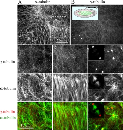
Figure 2.
Drosophila embryos lack a γ-tubulin–containing MTOC during interphase. An embryo in the process of dorsal closure was fixed and stained for α-tubulin (A) and γ-tubulin (B). Inset shows embryo model with red box showing relative orientation of images in A and B. Three different cell populations are boxed in A, corresponding to G2-arrested amnioserosa cells (1), G1-arrested leading-edge cells (2) and epithelial cells undergoing mitosis (3). Boxed regions are displayed at higher magnification. Insets show a bipolar spindle from a mitotic domain of a younger developing embryo with γ-tubulin labeling at the poles.

