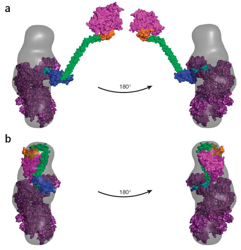
Figure 5.

SAXS envelope and models for full-length myosin VI. (a) A model of an extended full-length M6 containing the published post-stroke crystal structure of the catalytic domain5 with the calmodulin-bound unique insert and IQ regions (purple), the PT-DT model from Figure 3b and the Rosetta prediction of the CBD (magenta) docked into the corresponding SAXS envelope. With the head aligned to one end of the reconstruction and the PT fused to the IQ such that it extends the lever arm in the same conformation as in Figure 6, the rest of the tail lies well outside the calculated scattering envelope. (b) A model of an alternate compact state for monomeric M6, with the CBD folded back onto the lever arm calmodulins, docked into the same SAXS envelope as in a.
