Figure 4.
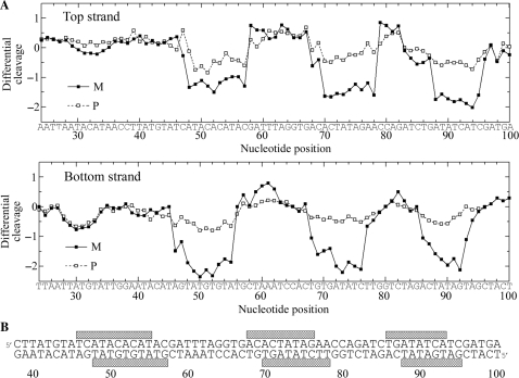
(A) Differential cleavage plots for M-[Fe2L3]4+ (full squares) and P-[Fe2L3]4+ (open squares) induced differences in susceptibility to DNase I digestion on top and bottom strands of the 158-mer HindIII/NdeI fragment of the plasmid pSP73 at 20:1 (base:cylinder) ratio. Vertical scales are in units of ln(fc) – ln(f0), where fc is the fractional cleavage at any bond in the presence of cylinder and f0 is the fractional cleavage of the same bond in the control, given closely similar extents of overall digestion. Positive values indicate enhancement, negative values inhibition. (B) Part of the sequence of 158-mer HindIII/NdeI fragment of the plasmid pSP73 showing preferential binding sites (shown as light bars) of M-[Fe2L3]4+ at 20:1 (base:cylinder) ratio. The binding sites were obtained by shifting the sites of inhibited DNase I cleavage by 2 bp in the 3′ direction.

