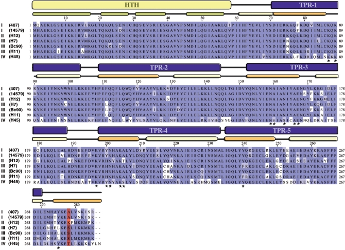
Figure 8.
Sequence alignment of seven representative PlcR sequences. Sequence alignment of seven representative PlcRs was performed with JalView, using the Blosum62 colouring scheme. The strain names are indicated in parentheses and the roman numbers refer to the PlcR groups. Based on 3D-structure, HTH domain and TRP domains are indicated in yellow and in blue, respectively. The α-helices from the HTH domain are in green. The first and the last α-helices from TPR domains are in light yellow and in orange, respectively. Asterisks represent PlcR residues implicated in PapR binding. Position 278 is coloured in red.

