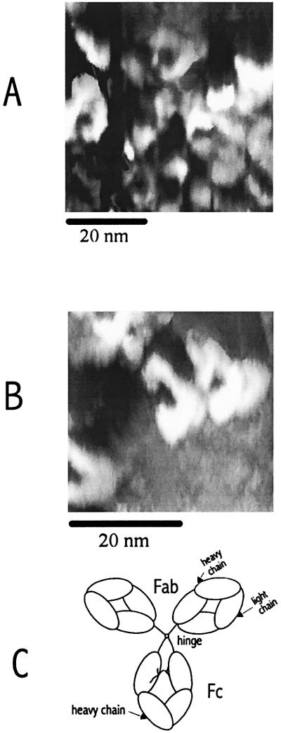
Figure 8.
(A and B) Constant-current images showing several mouse monoclonal IgG molecules on mica substrates treated with phosphate buffer. The images were recorded with a W tip in humid air (80% RH at 25°C). The reference current was 1 pA at a tip bias of 1.5 V (vs. the Au contact). (C) Schematic of an IgG molecule showing two Fab arms and the Fc portion (16).

