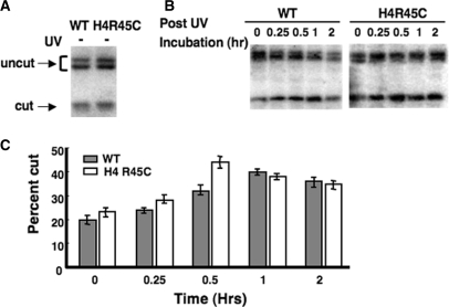
Figure 4.
Accessibility of the EcoRV site in HML chromatin. Nuclei were isolated from wt and H4 R45C cells (A) before or (B) after UV irradiation (100 J/m2) and digested with EcoRV, as described in Materials and methods section. Arrows show the positions of the uncut and cut bands. (C) Percent of HML DNA in chromatin accessible to EcoRV, where bars show values obtained for wt (shaded) and H4 R45C (open) cells. Data represent the mean ± 1 SD of three independent experiments.

