
Fig. 1. Mechanism and structure of IMPDH.
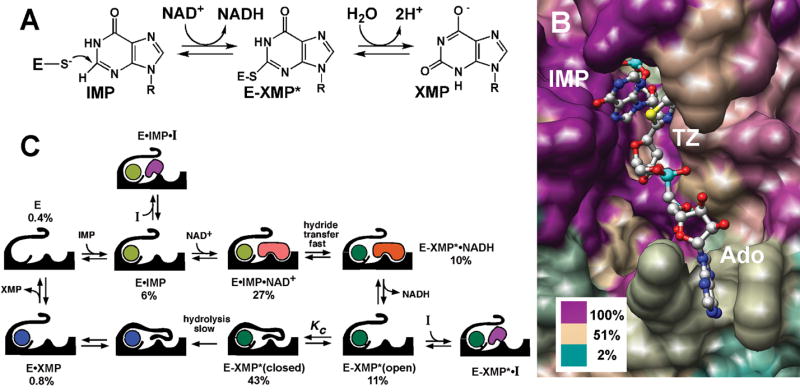
(A) The IMPDH reaction. (B) The active site, rendered by sequence conservation. The structure of E•IMP•tiazofurin adenine dinucleotide complex of IMPDH from Tritrichomonas foetus (PDB accession code 1LRT). Image was produced using the UCSF Chimera package (Pettersen et al., 2004). Percentage of sequence identity is colored as shown using the alignment from (Striepen et al., 2002). TZ, tiazofurin; Ado, adenosine. (C) The kinetic mechanism of IMPDH, showing the distribution of enzyme under the conditions of the HTS (250 μM IMP and 500 μM NAD+), determined as described in Supplemental Material. Not shown: E•NAD+ ≤ 0.7%, E-XMP*•NAD+ = 1%.