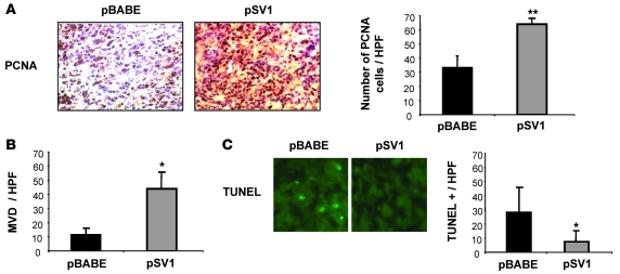
Figure 5. Changes in KLF6-SV1 metastatic tumor behavior correlates with markers of cellular proliferation, angiogenesis, and apoptosis.
Metastatic tumors were analyzed for their expression of PCNA, CD31, and TUNEL. (A) Immunohistochemistry for PCNA in pBABE vector– and KLF6-SV1–derived tumors. KLF6-SV1 caused increased PCNA staining in vivo. Eight independent high-power fields for each pBABE (n = 4) and KLF6-SV1 (n = 6) tumor were counted, assessing both the total number of cells and PCNA-positive cells. The graph represents the average percentage of PCNA-positive cells for each group. (B) CD31 staining of pBABE and KLF6-SV1 cell line–derived tumors. Overexpression of KLF6-SV1 protein caused a 4-fold increase in microvessel density (MVD), as measured by the number of CD31-positive endothelial cells per ×400 high-power field (HPF). (C) TUNEL staining of pBABE and KLF6-SV1 tumors. Representative images are shown. Overexpression of KLF6-SV1 decreased apoptosis by 70%. For each tumor, 6 high-power fields were counted and the total number of TUNEL-positive cells was determined. *P < 0.01, **P < 0.001 versus control. Original magnification, ×400.

