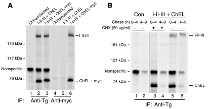
Figure 6. Secreted I-II-III protein is physically associated with secretory ChEL protein.
(A) 293 cells were transiently transfected with 0.5 μg plasmid DNA encoding I-II-III and cotransfected with 2.5 μg of plasmid DNA encoding the secretory ChEL domain either lacking or containing a myc epitope tag, as indicated. The cotransfected cells or untransfected controls were pulse labeled for 30 minutes and chased in complete media, and the secretion after 6 hours was analyzed by immunoprecipitation with anti-Tg or anti-myc. Immunoprecipitates and coprecipitates were analyzed by reducing 5.5% SDS-PAGE and fluorography. Addition of the myc tag slightly retards the SDS-PAGE mobility of the ChEL domain. Note that anti-myc precipitation of ChEL-myc coprecipitates I-II-III. (B) Cells untransfected (control) or cotransfected and pulse labeled as in A (I-II-III + ChEL) were chased in complete media for the time intervals shown, in the presence or absence of cycloheximide (CHX). The media were immunoprecipitated with anti-Tg and analyzed by reducing SDS-PAGE and fluorography. These lanes were run contiguously; a black line has been added for clarity to separate the samples. Note that prelabeled ChEL secretion proceeded rapidly in the presence of CHX, but prelabeled I-II-III secretion was blocked. The positions of molecular mass markers are shown at left.

