
Figure 5.
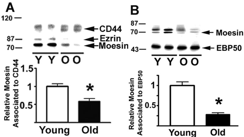
Age-related declines in the association of ERM to CD44 and EBP50. CD4 T cells from young and old CB6F1 donors were lysed, CD44 or EBP50 immunoprecipitated, and the associated ERM, CD44 and ERBP50 quantified by western blots. Panel A shows ERM associated with CD44; the bar plot shows mean and SEM of 13 young and 19 old mice tested in 4 experiments. Panel B shows the ERM levels immunoprecipitated by antibody to EBP50; the plot shows mean and SEM of 8 young and 14 old mice tested in 4 independent experiments. (*) indicates statistically significant effects.