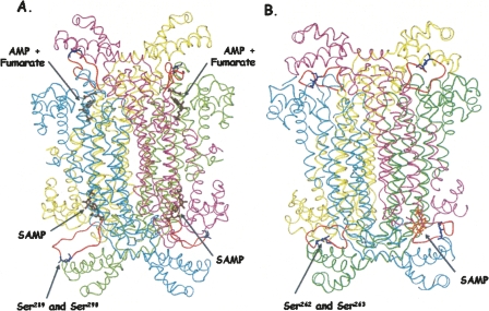
Figure 6.
Human and B. subtilis ASL. (A) Human ASL (PDB# 2VD6), with the loop region modeled in. SAMP and AMP + fumarate are pictured in brown. (B) The B. subtilis ASL model was based on the T. maritima ASL structure (PDB# 1C3U) with the SAMP docked in one active site (shown in orange). The serine residues in human (S289 and S290) and B. subtilis (S262 and S263) are shown in blue on the orange loops. The active site in the lower left of panel A is enlarged in Figure 8.

