
Figure 1.
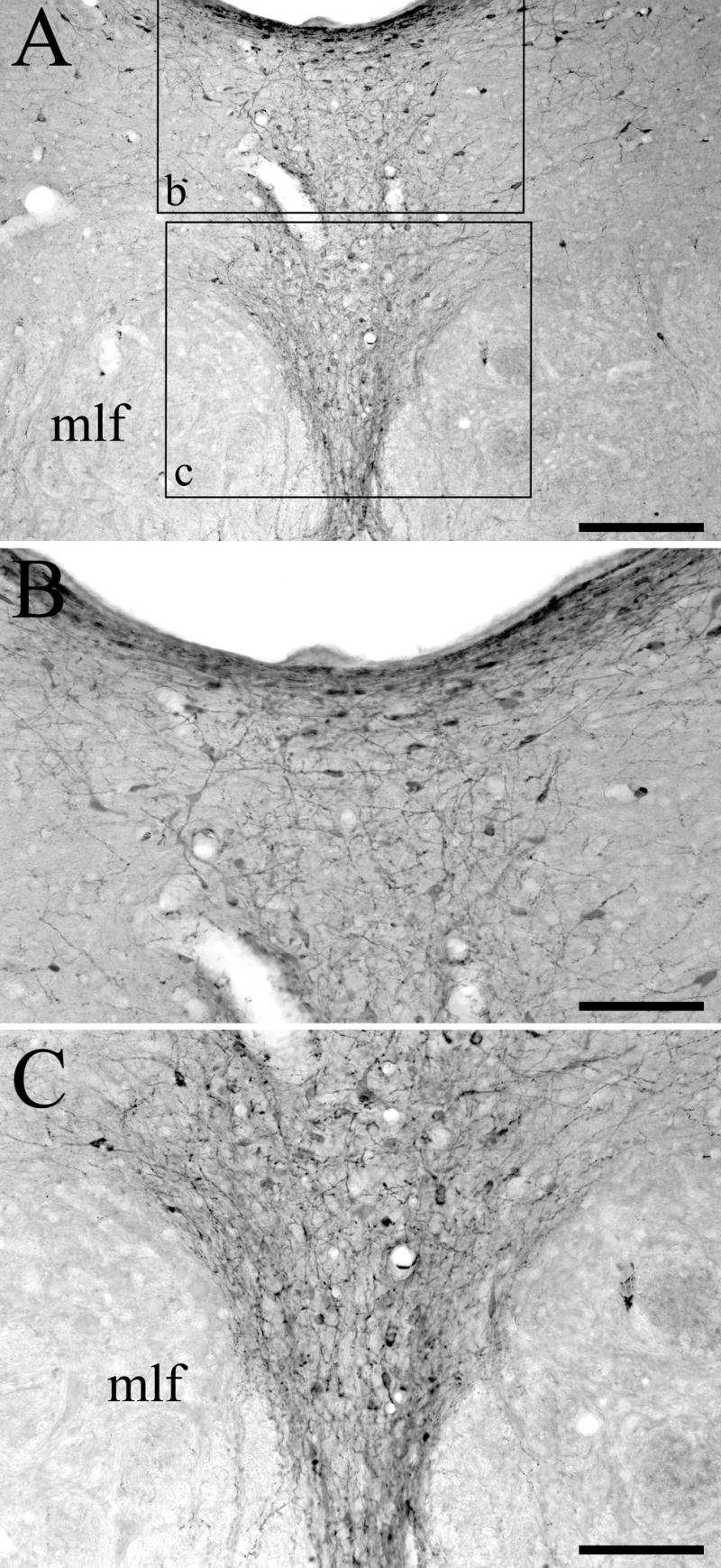
Tyrosine hydroxylase (TH) immunoreactivity in the DRN of animals treated with i.c.v. 5,7-DHT. (A) Numerous TH-positive cell bodies, dendrites, and fibers are located throughout the DRN. The boxes in panel A indicate the areas shown enlarged in panels B and C. (B) High-power photomicrograph showing TH immunostaining in the dorsomedial DRN subdivision. (C) High-power photomicrograph showing TH immunostaining in the ventromedial DRN subdivision. The medial longitudinal fasciculus (mlf) is also indicated. Scale bars = 200 μm in A and 100 μm in B and C.